

Swelling can change the tissues beneath the skin long before those changes are fully visible from the outside. While physical examination and medical history remain central to diagnosing lymphatic and venous conditions, ultrasound can provide additional information. This noninvasive imaging technology helps physicians examine tissue composition, fluid accumulation, fibrosis, and certain circulation problems that may contribute to persistent swelling.
Dr. Emily Iker has helped advance the clinical use of diagnostic ultrasonography in lymphatic medicine through pioneering published research. At the Lymphedema Center in Santa Monica, she combines ultrasound findings with a detailed evaluation of each patient’s symptoms, tissue changes, medical history, and swelling pattern. This integrated approach helps distinguish among conditions that may appear similar while supporting more individualized recommendations.
What Is Diagnostic Ultrasound?
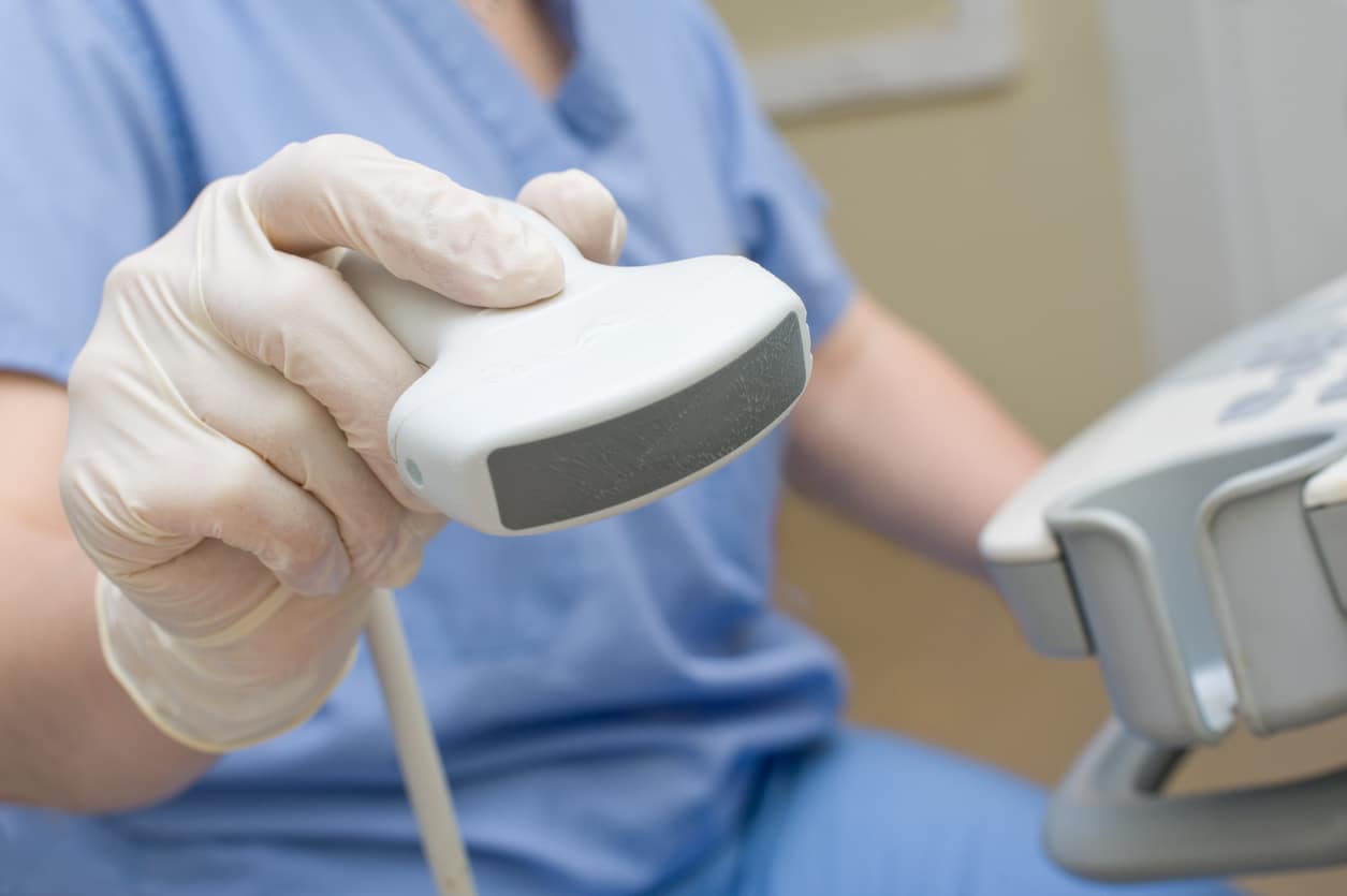
Diagnostic ultrasound, also called ultrasonography, uses high-frequency sound waves to create images of structures beneath the skin. A small handheld device called a transducer is moved across the area being examined. The sound waves reflect differently as they encounter fluid, fatty tissue, connective tissue, blood vessels, and other structures.
Ultrasound does not use radiation and does not require an incision. Most examinations are comfortable, although patients with very tender tissue may notice mild pressure as the transducer moves over the affected area.
Different types of ultrasound answer different clinical questions. Tissue ultrasonography may be used to examine changes associated with lymphatic or adipose tissue disorders. Vascular ultrasound focuses on blood flow through the veins or arteries. Depending on the symptoms, a patient may need one type of assessment or coordinated testing involving both.
How Does Ultrasound Support Lymphatic Assessment?
When lymphatic drainage is impaired, protein-rich fluid can accumulate within the tissues. Over time, ongoing inflammation may contribute to thickening and fibrosis. These changes can alter the appearance of the skin and subcutaneous tissues on ultrasound.
Diagnostic ultrasonography may help Dr. Iker assess:
-
Skin and subcutaneous tissue thickness
-
Patterns of fluid accumulation
-
Fibrosis or tissue hardening
-
Differences between affected and unaffected areas
-
Changes that may not be reflected by circumference measurements alone
-
Tissue characteristics associated with lymphedema or lipedema
-
Progression or improvement documented during follow-up care
Research continues to evaluate how ultrasound measurements can support earlier and more objective assessment of lymphedema. Recent clinical evidence indicates that ultrasound-based skin measurements may reveal changes that limb circumference alone does not capture, although ultrasound is not a universal stand-alone diagnostic test.
Can Ultrasound Help Distinguish Different Swelling Conditions?
Several conditions can enlarge an arm or leg, yet they do not produce identical changes beneath the skin. Ultrasonography may provide useful supporting information when distinguishing among:
-
Lymphedema
-
Lipedema
-
Lipo-lymphedema
-
Phlebo-lymphedema
-
Generalized fluid retention
-
Localized fatty tissue or lipomas
-
Inflammatory tissue changes
For example, lymphedema may lead to fluid accumulation and progressive fibrosis, while lipedema primarily affects the subcutaneous fat and connective tissue in a characteristic distribution. Some patients have overlapping conditions, so imaging findings must be interpreted alongside symptoms and physical signs.
Ultrasound cannot independently determine the cause of every type of swelling. Bloodwork, lymphoscintigraphy, MRI, CT imaging, genetic evaluation, or consultation with another medical specialist may be recommended when additional information is needed.
What Is the Role of Venous Ultrasound?
Venous disease can cause or worsen swelling, particularly in the lower legs. A venous duplex ultrasound combines traditional imaging with Doppler technology to evaluate the direction and speed of blood flow. It can help identify blood clots, venous obstruction, or valves that allow blood to flow backward.
This testing differs from the tissue ultrasonography used to assess changes associated with lymphatic dysfunction. When symptoms suggest chronic venous insufficiency, a previous deep vein thrombosis, or another vascular problem, Dr. Iker may recommend venous testing through an appropriate vascular provider.
Identifying venous involvement is especially important in suspected phlebo-lymphedema. Longstanding venous pressure can place increasing demands on the lymphatic system until both systems contribute to persistent swelling. In these cases, care may need to address the lymphatic component while a vascular specialist evaluates or treats the venous condition.
How Can Ultrasound Guide Ongoing Care?
Ultrasound assessment can help document a baseline before treatment or long-term management begins. Follow-up imaging may then show whether tissue thickness, fluid accumulation, or fibrosis has changed over time.
This information may help shape recommendations involving manual lymphatic drainage, movement, skin care, pneumatic compression, or properly selected compression garments. When compression is appropriate, options may include the practice’s FDA-approved compression sleeve design. Treatment decisions also account for the patient’s circulation, comfort, mobility, health risks, and ability to maintain a home routine.
Why Choose Dr. Emily Iker for Diagnostic Ultrasonography?
Ultrasound findings are most useful when interpreted by a physician who understands the varied ways in which lymphatic, venous, and adipose disorders affect tissues. Dr. Iker has dedicated more than three decades to evaluating and managing these complex conditions. Her clinical experience enables her to link imaging findings to subtle differences in swelling distribution, texture, symptoms, and function.
The Lymphedema Center is an LE&RN-designated Center of Excellence focused on lymphatic disorders. Rather than applying the same testing process to every patient, Dr. Iker selects and interprets diagnostic studies according to the individual presentation. When venous studies or additional imaging are needed, she can recommend appropriate referrals and help patients understand how those results relate to their lymphatic care.
Schedule an Ultrasound Assessment
Diagnostic ultrasonography can provide valuable information about tissue changes and the possible lymphatic or venous factors contributing to swelling. Call Lymphedema Center – Emily Iker, MD at (310) 829-7472 to schedule a consultation for an ultrasound assessment in Santa Monica, CA.